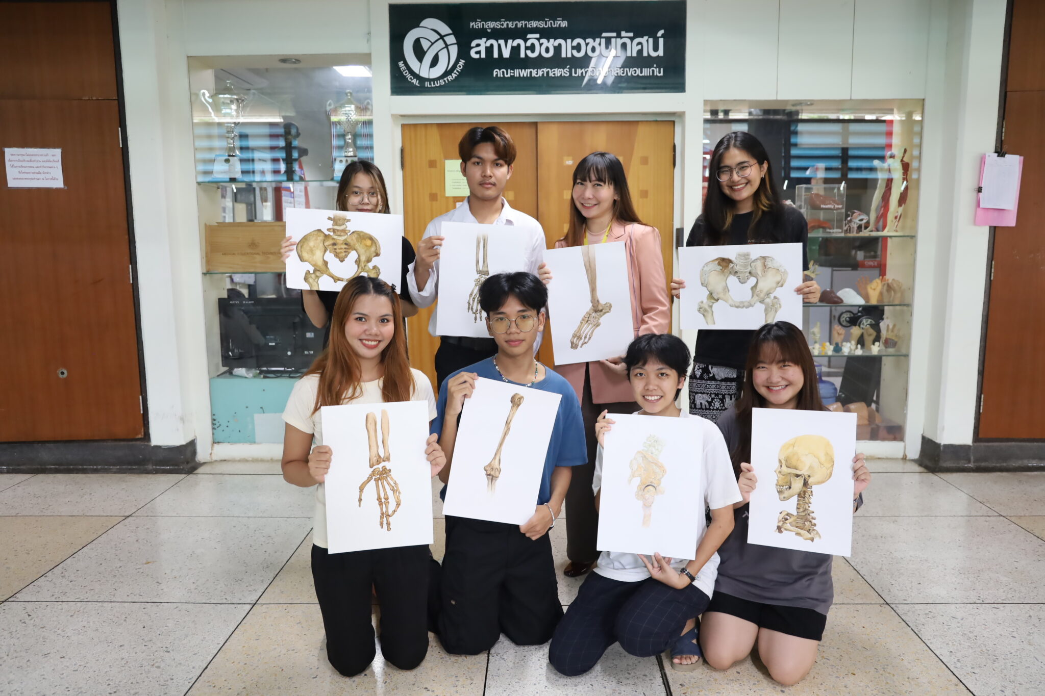
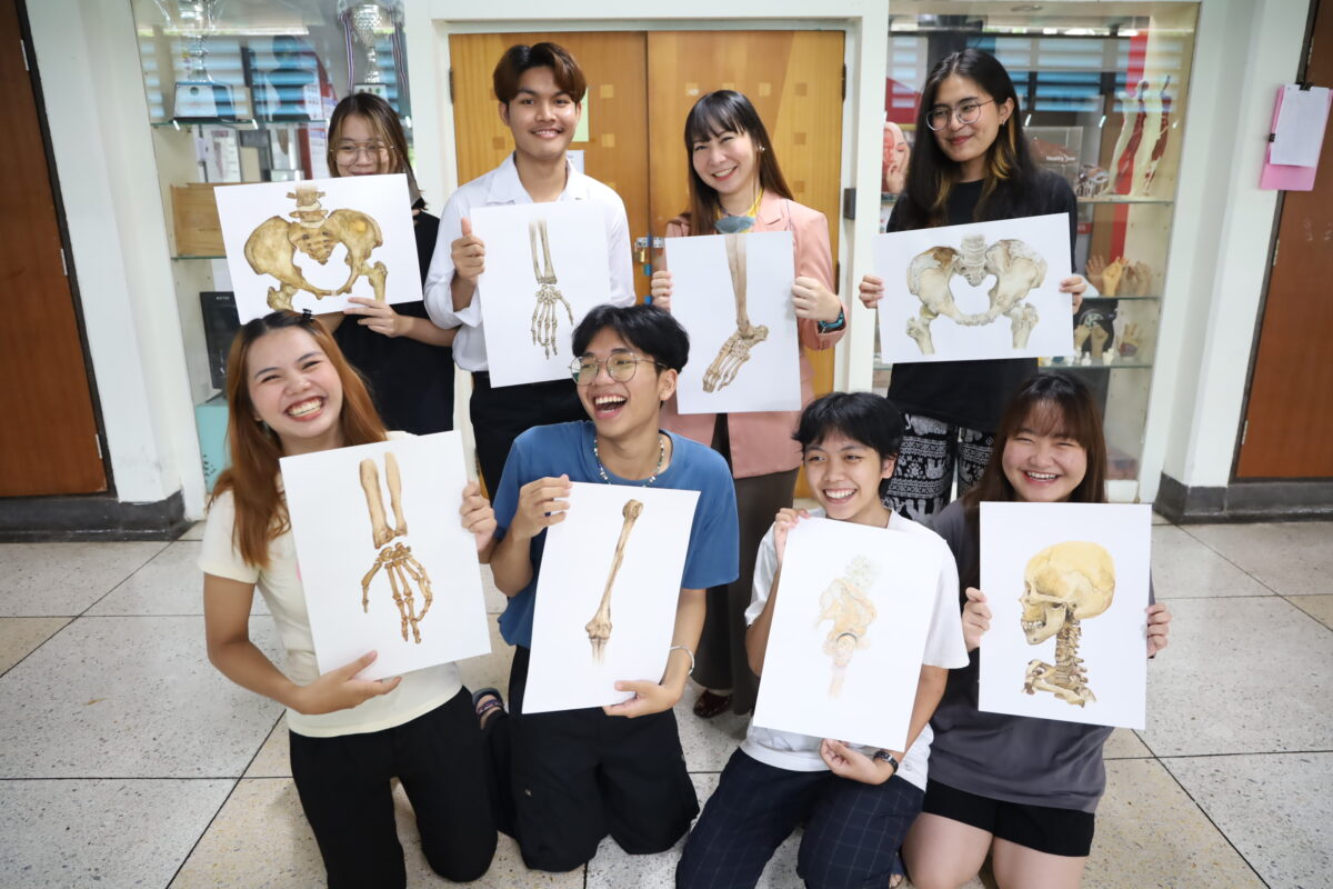
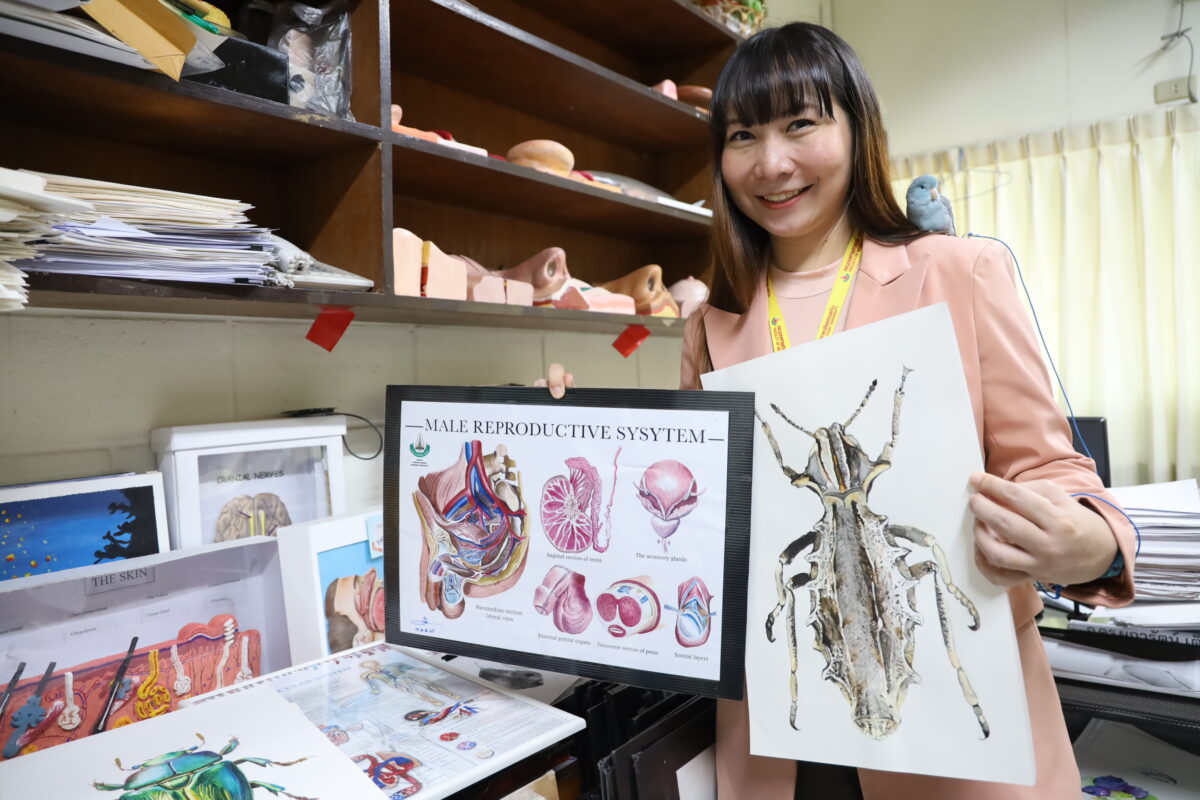
Viral has been ongoing on online social media after Facebook of “Chommanat Uppachitkul”, a lecturer of the Medical Illustration Program, Faculty of Medicine, Khon Kaen University posted the water-color paintings of Ajarn Yais’ (whole body donors’) bones, done by 2nd year students of the program. All paintings look so real like photographs that a lot of netizens made a lot of appreciative comments.
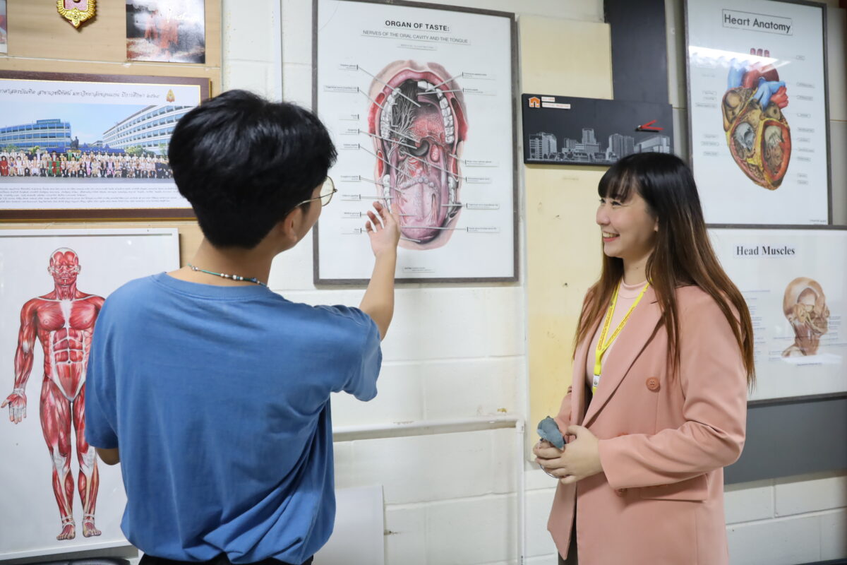
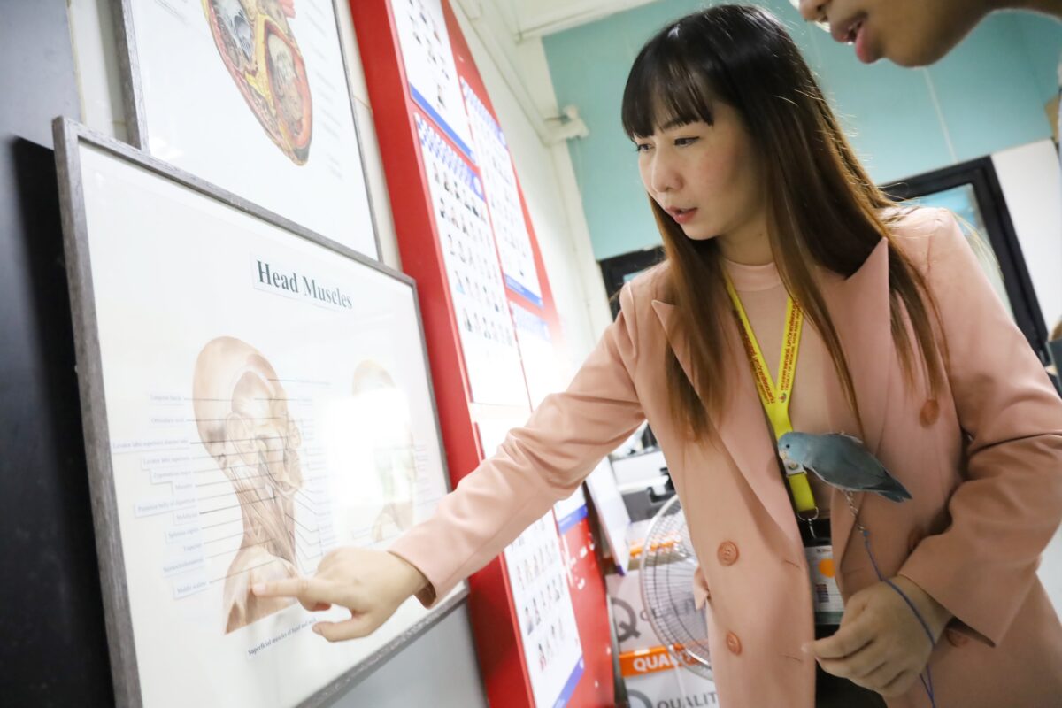
These works are part of the course, Advanced Painting for Medical Illustration, which is under the Medical Illustration Program of the Faculty of Medicine. “Even though the students are studying in the Faculty of Medicine, these students are not becoming doctors,” said Dr. Chommanat, “Medical Illustration Branch teaches students to construct medical media including printing items, models, video clips and photographs, which are used in the study of medicine. They even learn to design applications that assist in communications of doctors, medicine students and patients for better understanding.”
Medical Illustration students are not doctors, they are science students with the heart of arts.
“Medical Illustration program is a four-year program. Students learn medical courses such as clinical symptoms, parasitology or Gross Anatomy from doctors in parallel with learning techniques for constructing technological and art media. We can define our students as science students with the heart of arts.”
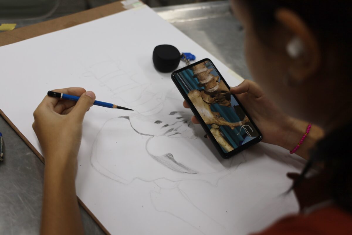
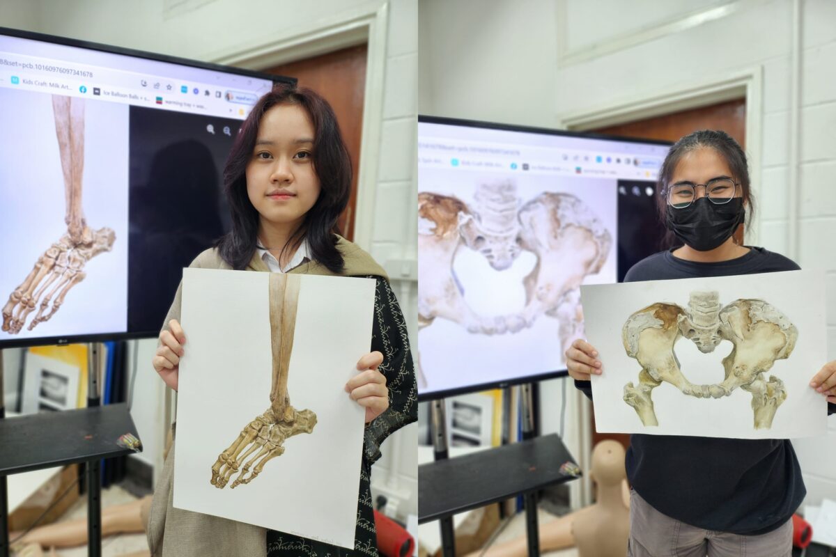
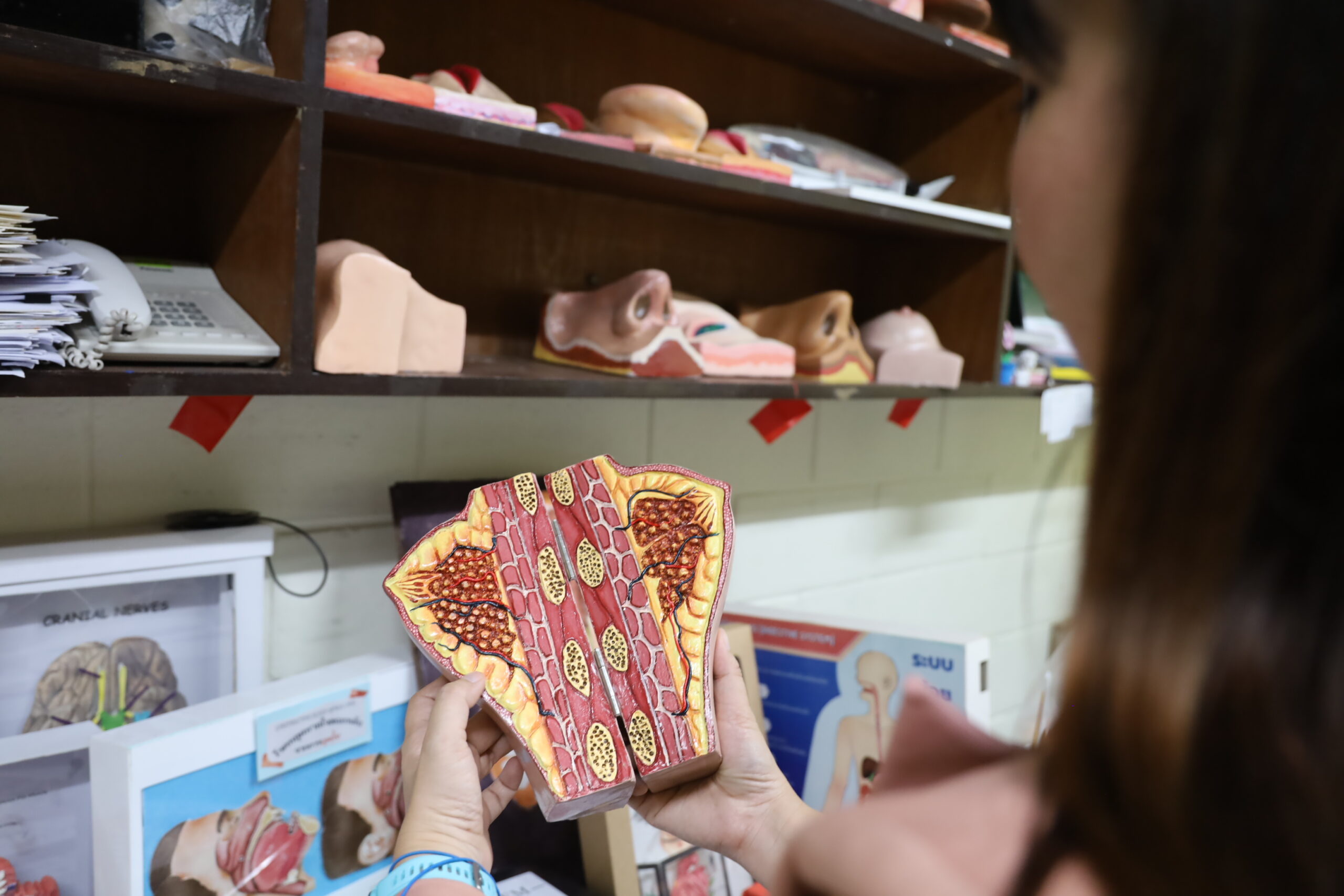
Nevertheless, medical-related drawing relies on different techniques from ordinary drawing. In order to see the reality, the drawing or painting must contain precise details so that they can be academically referred to. Before a piece of drawing or painting can be used, it must be checked by an expert or a doctor. It is not only a beautiful piece of work!
With intention to mentally support the students and to compile their works, Dr. Chommanat decided to post the paintings on social media. It turned out that the results are unexpected. The total ‘likes’ and ‘shares’ continuously increase. Meanwhile, doctor lecturers or lecturers from other faculties have contacted her, asking for the students to help drawing different medical science pictures for use as teaching media.”
First practice drawing of body donors’ bones
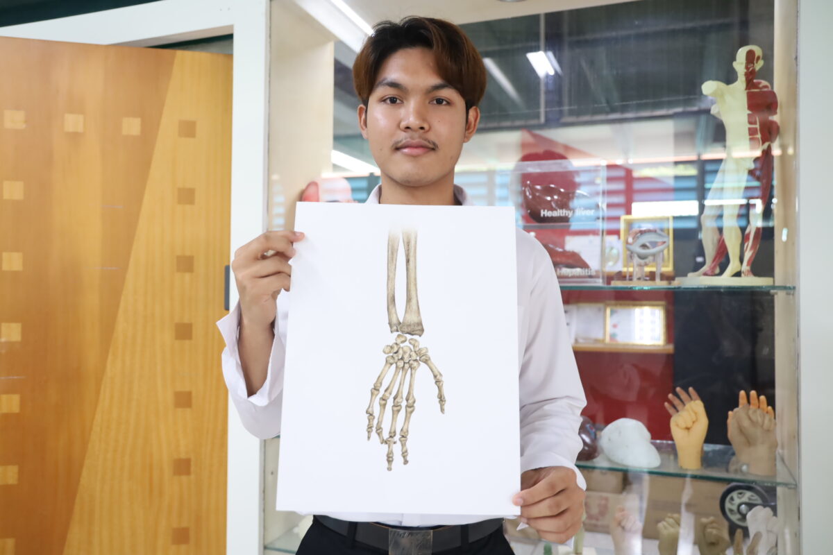
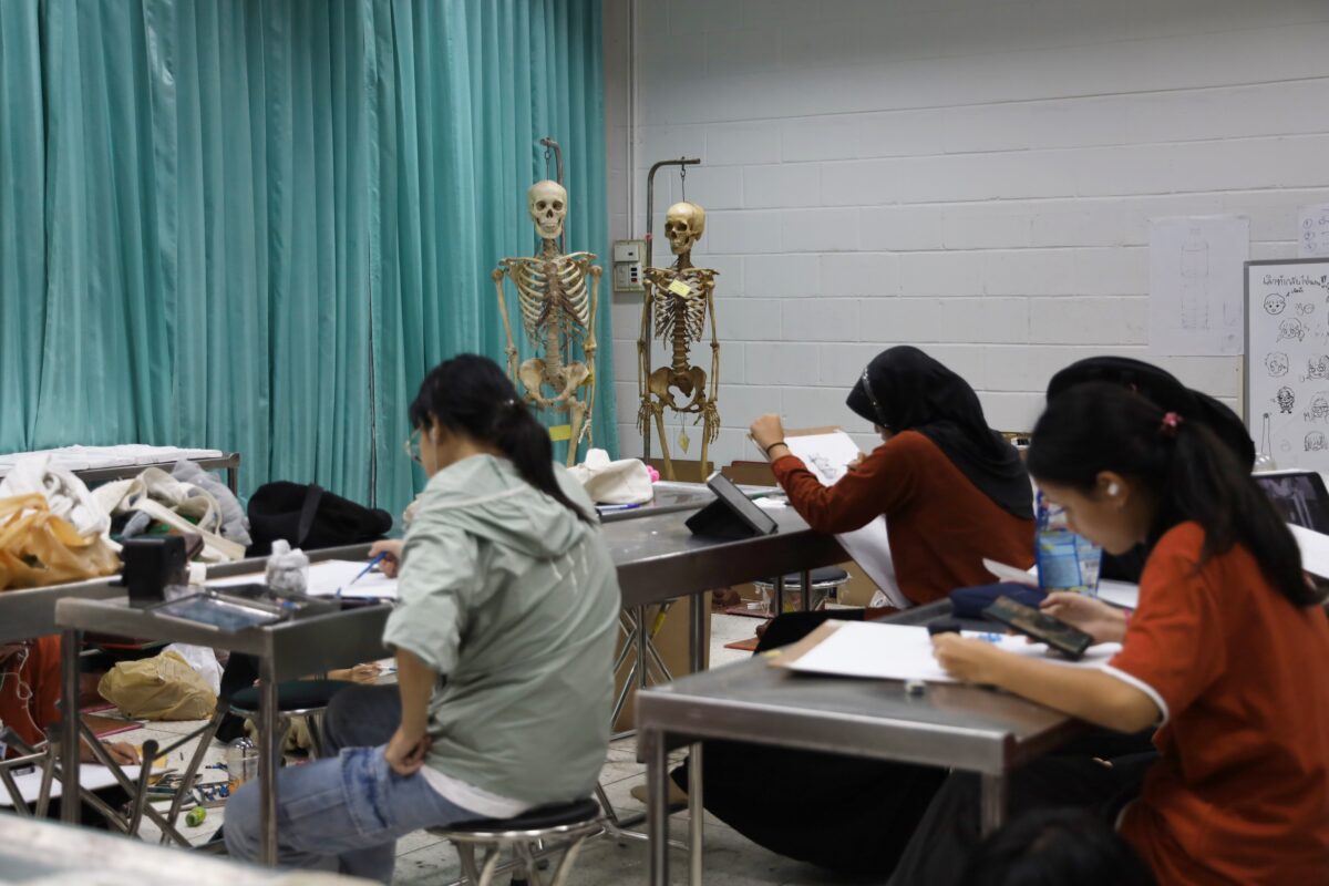
Mr. Nontakorn Janwan (Peem), a second year Medical Illustration student of the Faculty of Medicine said that after he had seen the feedback on social media, he was very glad that there are people appreciating the work. It was the first time the students drew and painted pictures of Ajarn Yais’ bones. And it was the first time he was using advanced water coloring technique. Before drawing, he had to measure each piece of bone, study the light angle and shade, then collected different details until he was sure the picture would resemble the reality and would be precise in all details. The end result is his painting of arm and hand bones.
“I chose arm and hand bones because they contain a lot of details. I spent 3-5 hours on these. The results are satisfactory. I hope the painting will be used as the learning media or educational media in medicine in the future.”
Although not knowing about Medical Illustration program before, Nontakorn had selected the program because he was interested in an integrated program of science and art. He hopes that his medical media construction will not only be used as the media in medical education, but will be a mediator between doctors and patients to communicate some difficult matters for better understanding.
Adding multi-skills for students for their future digital job market
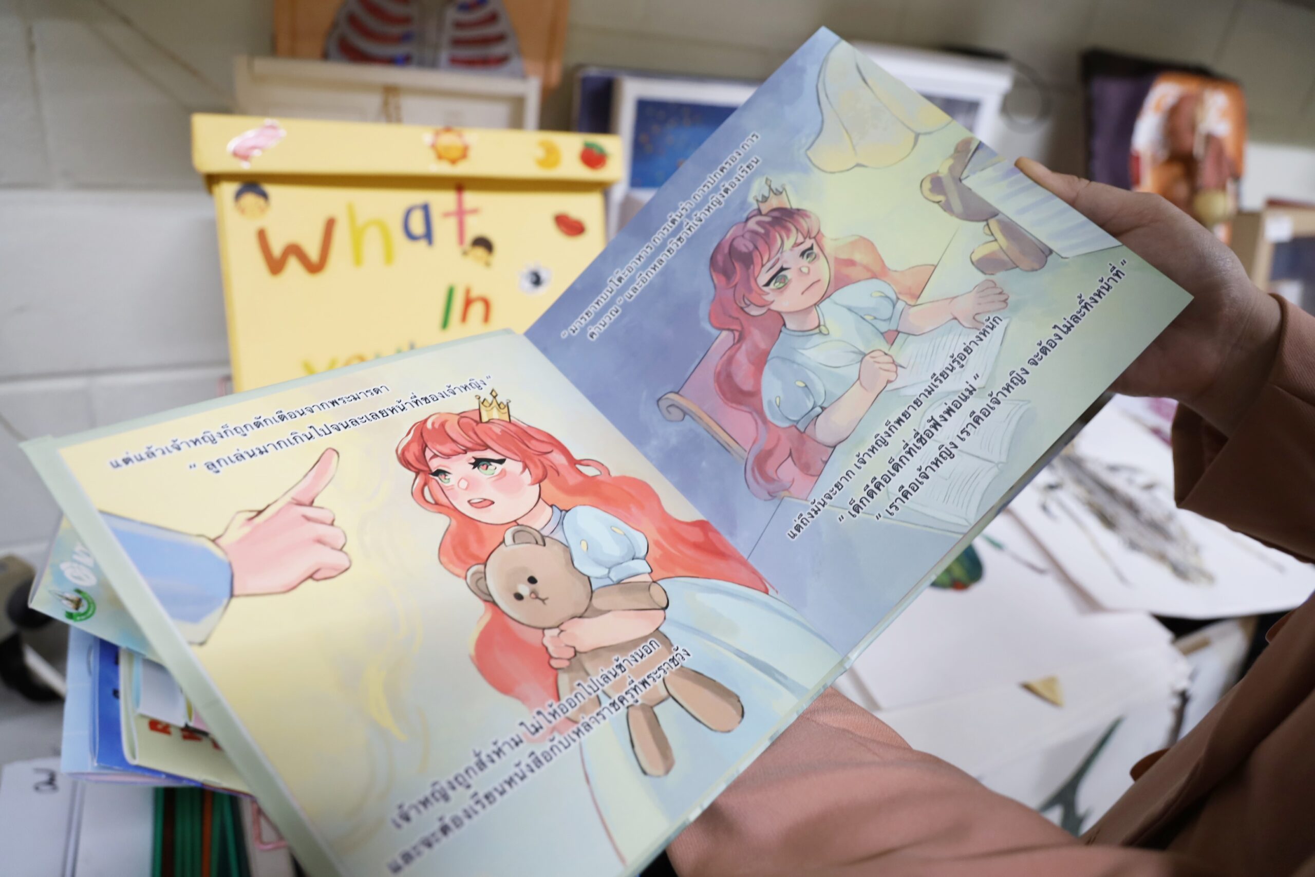
Dr. Chommanat added that for those interested to continue their studies in Medical Illustration program, they do not need to be able to draw well or like both science and arts. The important thing is the student should like to learn through real practices. During the first and second years of the program, students will learn about communication for medical illustration through practices of drawing, basic arts, graphics and sculpturing.
When they are in their 3rd year, students begin to learn the course on Medical Models, Practice of Making Motion Graphic, Coding, Cartoon Drawing for Medical Illustration and Medical Photography. These are multiple skills before they become a 4th year student when they will learn the course on Exhibition, Medical Media Project in which students are able to select the item they like and are interested in. The project can be done with a lecturer of a student from another faculty in order to integrate different sciences for students to learn the context of the work of Medical Illustration officers in hospitals and construction of teaching and learning media in the field.
“We have more students applying to the program now. We have always developed and revised the program by incorporating modern technologies for students to practice. When each student did his or her cooperative education program, the workplace became interested in each student and over 30% recruited them as employees, both in Thailand and in ASEAN countries. It has been found that the unemployment rate among our students is low and there are various jobs available. For more information, visit Facebook Page of the Medical Illustration Program, Faculty of Medicine, Khon Kaen University.”
News / Photos: Panit Khatnak